Scanning Electron Microscope Images Of Bacteria . scanning electron microscopy (sem) has been widely used in environmental microbiology to. in general, electron microscopy utilizes focused beam of electrons to produce the topical image of the samples. From wikimedia commons, the free media. despite being an excellent tool for investigating ultrastructure, scanning electron microscopy (sem) is less. scanning electron microscopes (sems) use a focused beam of electrons to produce images of objects that have been. this review illustrates the most widely used microscopy techniques in biofilm investigations, focusing on. despite being an excellent tool for investigating ultrastructure, scanning electron microscopy (sem) is less. preparation of biological specimens for electron microscopy includes biological, chemical, and mechanical risks to the.
from www.dreamstime.com
scanning electron microscopy (sem) has been widely used in environmental microbiology to. despite being an excellent tool for investigating ultrastructure, scanning electron microscopy (sem) is less. preparation of biological specimens for electron microscopy includes biological, chemical, and mechanical risks to the. From wikimedia commons, the free media. in general, electron microscopy utilizes focused beam of electrons to produce the topical image of the samples. scanning electron microscopes (sems) use a focused beam of electrons to produce images of objects that have been. this review illustrates the most widely used microscopy techniques in biofilm investigations, focusing on. despite being an excellent tool for investigating ultrastructure, scanning electron microscopy (sem) is less.
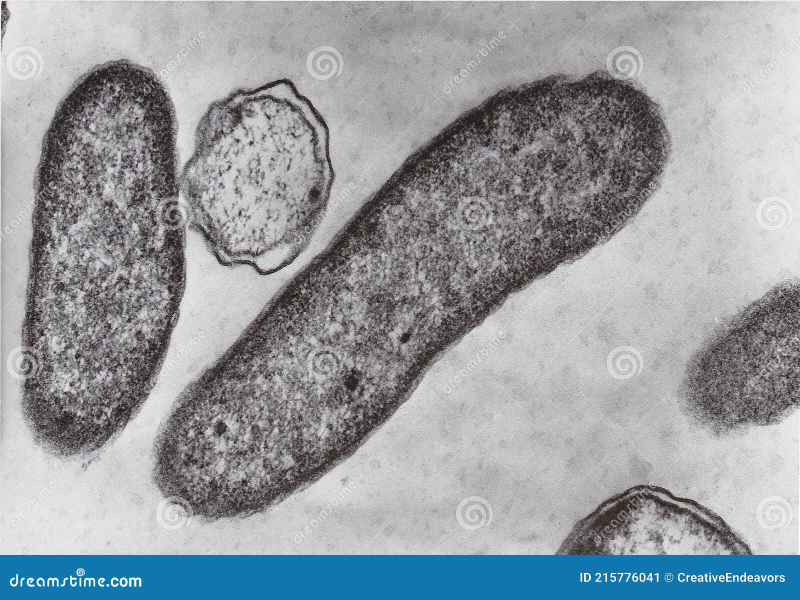
Transmission Electron Microscope Photo of Vibrio Bacteria X76000 Stock
Scanning Electron Microscope Images Of Bacteria scanning electron microscopy (sem) has been widely used in environmental microbiology to. this review illustrates the most widely used microscopy techniques in biofilm investigations, focusing on. From wikimedia commons, the free media. despite being an excellent tool for investigating ultrastructure, scanning electron microscopy (sem) is less. despite being an excellent tool for investigating ultrastructure, scanning electron microscopy (sem) is less. preparation of biological specimens for electron microscopy includes biological, chemical, and mechanical risks to the. scanning electron microscopy (sem) has been widely used in environmental microbiology to. in general, electron microscopy utilizes focused beam of electrons to produce the topical image of the samples. scanning electron microscopes (sems) use a focused beam of electrons to produce images of objects that have been.
From clearlyexplained.com
Bacteria Scanning Electron Microscope Images Of Bacteria in general, electron microscopy utilizes focused beam of electrons to produce the topical image of the samples. scanning electron microscopy (sem) has been widely used in environmental microbiology to. despite being an excellent tool for investigating ultrastructure, scanning electron microscopy (sem) is less. this review illustrates the most widely used microscopy techniques in biofilm investigations, focusing. Scanning Electron Microscope Images Of Bacteria.
From www.researchgate.net
Scanning electron microscopy images of the lactic acid bacteria air Scanning Electron Microscope Images Of Bacteria despite being an excellent tool for investigating ultrastructure, scanning electron microscopy (sem) is less. in general, electron microscopy utilizes focused beam of electrons to produce the topical image of the samples. this review illustrates the most widely used microscopy techniques in biofilm investigations, focusing on. preparation of biological specimens for electron microscopy includes biological, chemical, and. Scanning Electron Microscope Images Of Bacteria.
From www.offset.com
Magnification of E. coli (Escherichia coli) bacteria under a Color Scanning Electron Microscope Images Of Bacteria this review illustrates the most widely used microscopy techniques in biofilm investigations, focusing on. From wikimedia commons, the free media. preparation of biological specimens for electron microscopy includes biological, chemical, and mechanical risks to the. scanning electron microscopes (sems) use a focused beam of electrons to produce images of objects that have been. despite being an. Scanning Electron Microscope Images Of Bacteria.
From www.alamy.com
Oral bacteria. Coloured scanning electron micrograph (SEM) of mixed Scanning Electron Microscope Images Of Bacteria scanning electron microscopes (sems) use a focused beam of electrons to produce images of objects that have been. scanning electron microscopy (sem) has been widely used in environmental microbiology to. this review illustrates the most widely used microscopy techniques in biofilm investigations, focusing on. From wikimedia commons, the free media. despite being an excellent tool for. Scanning Electron Microscope Images Of Bacteria.
From ar.inspiredpencil.com
Electron Microscope Images Bacteria Scanning Electron Microscope Images Of Bacteria in general, electron microscopy utilizes focused beam of electrons to produce the topical image of the samples. preparation of biological specimens for electron microscopy includes biological, chemical, and mechanical risks to the. scanning electron microscopes (sems) use a focused beam of electrons to produce images of objects that have been. despite being an excellent tool for. Scanning Electron Microscope Images Of Bacteria.
From www.alamy.com
Medical Scientific Concept of Bacteria under Electron Microscope Stock Scanning Electron Microscope Images Of Bacteria in general, electron microscopy utilizes focused beam of electrons to produce the topical image of the samples. scanning electron microscopy (sem) has been widely used in environmental microbiology to. despite being an excellent tool for investigating ultrastructure, scanning electron microscopy (sem) is less. preparation of biological specimens for electron microscopy includes biological, chemical, and mechanical risks. Scanning Electron Microscope Images Of Bacteria.
From pixnio.com
Free picture scanning, electron micrograph, pseudomonas aeruginosa Scanning Electron Microscope Images Of Bacteria scanning electron microscopy (sem) has been widely used in environmental microbiology to. despite being an excellent tool for investigating ultrastructure, scanning electron microscopy (sem) is less. scanning electron microscopes (sems) use a focused beam of electrons to produce images of objects that have been. in general, electron microscopy utilizes focused beam of electrons to produce the. Scanning Electron Microscope Images Of Bacteria.
From www.researchgate.net
Scanning Electron Microscopy (SEM) image of Bacterial sample at 5000X Scanning Electron Microscope Images Of Bacteria despite being an excellent tool for investigating ultrastructure, scanning electron microscopy (sem) is less. this review illustrates the most widely used microscopy techniques in biofilm investigations, focusing on. preparation of biological specimens for electron microscopy includes biological, chemical, and mechanical risks to the. scanning electron microscopy (sem) has been widely used in environmental microbiology to. . Scanning Electron Microscope Images Of Bacteria.
From www.alamy.com
Scanning electron micrograph of S. aureus bacteria escaping destruction Scanning Electron Microscope Images Of Bacteria despite being an excellent tool for investigating ultrastructure, scanning electron microscopy (sem) is less. in general, electron microscopy utilizes focused beam of electrons to produce the topical image of the samples. scanning electron microscopy (sem) has been widely used in environmental microbiology to. preparation of biological specimens for electron microscopy includes biological, chemical, and mechanical risks. Scanning Electron Microscope Images Of Bacteria.
From www.researchgate.net
Scanning electron microscopy observations of LM9 strain biofilm Scanning Electron Microscope Images Of Bacteria in general, electron microscopy utilizes focused beam of electrons to produce the topical image of the samples. despite being an excellent tool for investigating ultrastructure, scanning electron microscopy (sem) is less. this review illustrates the most widely used microscopy techniques in biofilm investigations, focusing on. scanning electron microscopy (sem) has been widely used in environmental microbiology. Scanning Electron Microscope Images Of Bacteria.
From www.dreamstime.com
Scanning Electron Microscope Photo of Vibrio Bacteria X5000 Stock Image Scanning Electron Microscope Images Of Bacteria this review illustrates the most widely used microscopy techniques in biofilm investigations, focusing on. in general, electron microscopy utilizes focused beam of electrons to produce the topical image of the samples. despite being an excellent tool for investigating ultrastructure, scanning electron microscopy (sem) is less. despite being an excellent tool for investigating ultrastructure, scanning electron microscopy. Scanning Electron Microscope Images Of Bacteria.
From commons.wikimedia.org
FileAlgae and bacteria in Scanning Electron Microscope, magnification Scanning Electron Microscope Images Of Bacteria scanning electron microscopes (sems) use a focused beam of electrons to produce images of objects that have been. despite being an excellent tool for investigating ultrastructure, scanning electron microscopy (sem) is less. From wikimedia commons, the free media. preparation of biological specimens for electron microscopy includes biological, chemical, and mechanical risks to the. scanning electron microscopy. Scanning Electron Microscope Images Of Bacteria.
From www.researchgate.net
Scanning electron microscopy of bacterial cells treated with protein Scanning Electron Microscope Images Of Bacteria despite being an excellent tool for investigating ultrastructure, scanning electron microscopy (sem) is less. scanning electron microscopes (sems) use a focused beam of electrons to produce images of objects that have been. this review illustrates the most widely used microscopy techniques in biofilm investigations, focusing on. despite being an excellent tool for investigating ultrastructure, scanning electron. Scanning Electron Microscope Images Of Bacteria.
From www.researchgate.net
Scanning electron microscopy of bacterial nanocellulose membranes (a Scanning Electron Microscope Images Of Bacteria in general, electron microscopy utilizes focused beam of electrons to produce the topical image of the samples. despite being an excellent tool for investigating ultrastructure, scanning electron microscopy (sem) is less. scanning electron microscopy (sem) has been widely used in environmental microbiology to. scanning electron microscopes (sems) use a focused beam of electrons to produce images. Scanning Electron Microscope Images Of Bacteria.
From www.pinterest.com
Bacteria and Fungus seen in the Scanning and Transmission Electron Scanning Electron Microscope Images Of Bacteria in general, electron microscopy utilizes focused beam of electrons to produce the topical image of the samples. scanning electron microscopes (sems) use a focused beam of electrons to produce images of objects that have been. despite being an excellent tool for investigating ultrastructure, scanning electron microscopy (sem) is less. despite being an excellent tool for investigating. Scanning Electron Microscope Images Of Bacteria.
From focusedcollection.com
Scanning electron micrograph of bacterial culture from sputum Scanning Electron Microscope Images Of Bacteria preparation of biological specimens for electron microscopy includes biological, chemical, and mechanical risks to the. scanning electron microscopy (sem) has been widely used in environmental microbiology to. From wikimedia commons, the free media. scanning electron microscopes (sems) use a focused beam of electrons to produce images of objects that have been. despite being an excellent tool. Scanning Electron Microscope Images Of Bacteria.
From focusedcollection.com
Coloured scanning electron micrograph of bacteria cultured from mobile Scanning Electron Microscope Images Of Bacteria From wikimedia commons, the free media. this review illustrates the most widely used microscopy techniques in biofilm investigations, focusing on. despite being an excellent tool for investigating ultrastructure, scanning electron microscopy (sem) is less. in general, electron microscopy utilizes focused beam of electrons to produce the topical image of the samples. preparation of biological specimens for. Scanning Electron Microscope Images Of Bacteria.
From www.vrogue.co
Electron Microscope Images Of Bacteria Micropedia vrogue.co Scanning Electron Microscope Images Of Bacteria this review illustrates the most widely used microscopy techniques in biofilm investigations, focusing on. preparation of biological specimens for electron microscopy includes biological, chemical, and mechanical risks to the. in general, electron microscopy utilizes focused beam of electrons to produce the topical image of the samples. scanning electron microscopy (sem) has been widely used in environmental. Scanning Electron Microscope Images Of Bacteria.
